So, you find yourself in a bit of an emergency situation, and the question of whether you’ll need X-rays taken during your visit to the doctor’s office is on your mind. Well, let’s get straight to the point – X-rays can be a critical tool for diagnosing and understanding certain injuries or conditions. Whether it’s a broken bone, potential internal damage, or even a foreign object lodged where it shouldn’t be, X-rays can provide valuable insights that help healthcare professionals determine the best course of action. In this article, we’ll explore the importance of X-rays during emergency visits and shed some light on what you can expect if you find yourself in need of this imaging technique.

Reasons for X-rays during an emergency visit
Assessing fractures and broken bones
One of the primary reasons for getting an X-ray during an emergency visit is to assess fractures and broken bones. X-rays are essential in determining the extent of the injury and identifying the exact location of the fracture or break. This information is crucial for planning the appropriate treatment, such as a cast or surgery, and ensuring proper alignment of the bones during the healing process.
Identifying dislocations
X-rays are also valuable in identifying dislocations. A dislocation occurs when the bones in a joint are forced out of their normal positions. X-rays can help healthcare providers visualize the displacement of the bones, which guides them in the reduction process. By seeing the precise location and severity of the dislocation, doctors can effectively manipulate the joint back into its proper alignment, reducing pain and preventing further damage.
Detecting foreign objects
During an emergency visit, X-rays can be extremely useful in detecting foreign objects that may have penetrated the body. Whether it’s a glass shard, a metal fragment, or any other foreign object, X-rays can provide a clear image of its location, size, and potential damage it has caused to surrounding tissues or organs. This information is crucial for deciding the appropriate course of action, such as removing the object surgically or monitoring its impact on the patient’s health.
Evaluating joint or bone infections
X-rays also play a significant role in evaluating joint or bone infections. Infections in these areas can lead to severe complications if not promptly diagnosed and treated. X-rays can assist healthcare providers in identifying signs of infection, such as changes in bone density, abnormal bone growth, or fluid accumulation in the joint. With this information, doctors can prescribe appropriate antibiotics or recommend surgical intervention to remove infected tissues and prevent further spread of the infection.
Benefits of X-rays in emergency situations
Quick and accurate diagnosis
One of the key benefits of X-rays in emergency situations is their ability to provide quick and accurate diagnoses. X-rays produce immediate images that allow healthcare providers to assess injuries or conditions accurately. This quick diagnosis enables doctors to initiate the most appropriate and timely treatment plan, reducing the potential for complications and promoting faster recovery.
Guiding treatment decisions
X-rays assist in guiding treatment decisions by providing vital information about the nature and severity of the injury or condition. Whether it’s determining the need for surgery, selecting the appropriate immobilization device, or deciding on the administration of medication, X-rays offer healthcare providers crucial insights into the best course of action for each individual patient. This guidance ensures that patients receive the most effective and tailored treatment for their specific needs.
Minimizing unnecessary procedures
X-rays help in minimizing unnecessary procedures, saving time and resources for both patients and healthcare providers. By providing clear images of the internal structures, X-rays can help rule out certain conditions or injuries that may have otherwise required additional diagnostic procedures. This minimization of unnecessary procedures not only enhances the efficiency of emergency care but also reduces patient discomfort and exposure to potentially harmful interventions.
X-ray procedure during an emergency visit
Preparing for the X-ray
Before undergoing an X-ray during an emergency visit, certain preparations are necessary. You may need to remove any jewelry, clothing, or objects that could interfere with the imaging process. It is important to inform the healthcare providers of any known allergies, especially to contrast dye, which may be used in some X-ray procedures. Additionally, if you are pregnant or suspect you may be pregnant, it is essential to inform the medical staff to ensure appropriate precautions are taken.
Positioning for the X-ray
Once prepared, positioning for the X-ray will be determined by the specific part of the body being imaged. The healthcare provider will guide you into the correct position, ensuring optimal visualization of the area of interest. It is important to follow their instructions carefully to obtain the most accurate images and facilitate an accurate diagnosis.
Protecting from radiation
During an X-ray, steps are taken to protect you from unnecessary radiation exposure. Your healthcare provider will use a lead apron and shield to cover areas not being imaged, such as the reproductive organs, to minimize radiation exposure to those regions. Additionally, the X-ray machine will be adjusted to deliver the lowest possible radiation dose while still achieving the necessary image quality.
Taking X-ray images
Once properly positioned and protected, the X-ray machine will be positioned accordingly to capture the necessary images. These images are taken quickly and painlessly. You may be required to hold your breath momentarily to reduce motion blur and ensure clarity of the images. After the X-rays are taken, they will be reviewed by a radiologist or healthcare provider who will interpret the images and relay the findings to the treating physician.
Risks associated with X-rays
Exposure to radiation
One of the primary risks associated with X-rays is exposure to radiation. While the radiation dose used in X-rays is generally considered safe, repeated or prolonged exposure can potentially increase the risk of developing cancer or other radiation-related complications. However, it’s essential to note that the benefits of using X-rays to diagnose and treat urgent medical conditions often outweigh the small risk of radiation exposure.
Potential harm to pregnant women
X-rays also pose potential harm to pregnant women and their developing fetuses. Ionizing radiation used in X-rays can potentially cause harm to the developing baby, particularly during the first trimester. It is crucial for pregnant women to inform healthcare providers about their pregnancy to ensure proper precautions and alternative imaging methods are used whenever possible.
Possible allergic reaction to contrast dye
When contrast dye is used during certain X-ray procedures, there is a risk of an allergic reaction. Allergic reactions to contrast dye are relatively rare but can range from mild symptoms such as itching and rash to more severe reactions like difficulty breathing or anaphylaxis. Informing healthcare providers about any known allergies beforehand is essential to prevent potential allergic reactions and ensure appropriate measures are taken to manage or avoid the use of contrast dye.

Safety protocols during X-rays
Use of lead apron and shield
During an X-ray, safety protocols are in place to protect you from unnecessary radiation exposure. The use of a lead apron and shield is critical to minimize radiation exposure to areas not being imaged, such as the reproductive organs. These protective measures ensure that the radiation is focused only on the targeted area and significantly reduce the potential risks associated with X-rays.
Limiting unnecessary exposure
Healthcare providers are trained to limit unnecessary exposure to radiation during X-rays. By using the lowest radiation dose necessary to achieve the required image quality, healthcare providers can minimize radiation exposure without compromising the effectiveness of the diagnostic process. Regular monitoring of radiation dosage and adherence to established safety guidelines help ensure that exposure is kept as low as reasonably achievable.
Alternative imaging options
In some cases, alternative imaging options may be available to avoid or minimize the use of X-rays. For example, ultrasound imaging, MRI scans, or CT scans may be utilized based on the specific clinical situation. These alternative imaging methods provide valuable diagnostic information without using ionizing radiation. However, it is important to note that each imaging modality has its limitations and may not always be a suitable substitute for X-rays.
Common concerns about X-rays
Effectiveness compared to other imaging techniques
One common concern about X-rays is their effectiveness compared to other imaging techniques. While X-rays are excellent for visualizing bones and identifying fractures, they may not provide as detailed information about soft tissues and organs as other imaging techniques, such as MRI or ultrasound. However, in emergency situations where immediate assessment and diagnosis are crucial, X-rays remain an important and valuable tool.
Costs and insurance coverage
Another concern for many individuals is the cost of X-rays and insurance coverage. The cost of X-rays can vary depending on factors such as the facility, location, and specific type of X-ray being performed. However, it is important to note that X-rays are often covered by health insurance, especially in emergency situations. It is advisable to consult with your insurance provider to understand the specific coverage and potential out-of-pocket expenses associated with X-ray procedures.
Minimizing radiation exposure
Minimizing radiation exposure is a common concern among patients undergoing X-rays. While it is true that X-rays use ionizing radiation, it is important to remember that radiation doses are kept as low as reasonably achievable and well within safety limits. The benefits of using X-rays to diagnose and treat urgent medical conditions typically outweigh the small potential risks associated with radiation exposure. However, if you have any specific concerns or questions, it is always advisable to discuss them with your healthcare provider.

Determining the need for X-rays
Clinical examination and medical history
Determining the need for X-rays during an emergency visit involves a thorough clinical examination and consideration of the patient’s medical history. The healthcare provider will assess the signs and symptoms, conduct a physical examination, and inquire about the circumstances of the injury or condition. This information, combined with the patient’s medical history, helps guide the decision-making process and determine whether an X-ray is necessary for further evaluation.
Healthcare provider’s assessment
The healthcare provider’s assessment plays a crucial role in determining the need for X-rays. Based on their clinical experience and knowledge, they evaluate the urgency and potential benefits of obtaining X-ray images. Factors such as the severity of the injury, risk of complications, and potential impact on treatment decisions are taken into account. The healthcare provider’s assessment helps ensure that X-rays are ordered when necessary, minimizing unnecessary exposure while maximizing the diagnostic value.
Signs and symptoms
Signs and symptoms exhibited by the patient are important indicators for the need for X-rays. Visible deformities, severe pain, inability to move a limb, swelling, bruising, or abnormal range of motion are all potential signs that an X-ray may be necessary to evaluate the injury or condition. Additionally, certain symptoms like persistent coughing, chest pain, neurological deficits, or abdominal tenderness may warrant X-rays to identify potential underlying causes. These signs and symptoms guide the healthcare provider in determining whether X-rays are essential for accurate diagnosis and appropriate treatment.
X-rays for specific emergency conditions
Head injuries and concussions
X-rays are commonly used to evaluate head injuries and concussions. While they may not always be the initial imaging choice, they can be valuable in detecting skull fractures, identifying the presence of foreign bodies, or ruling out severe brain trauma. X-rays can provide important information that helps healthcare providers determine the best course of action to manage head injuries and ensure patient safety.
Chest and lung injuries
X-rays are frequently utilized to assess chest and lung injuries during emergency visits. They can quickly identify fractures of the ribs or sternum, detect collapsed lungs (pneumothorax), or assess the severity of lung infections, such as pneumonia. X-rays can also reveal other abnormalities, including fluid accumulation or foreign objects in the chest, aiding in the diagnosis and subsequent treatment decisions.
Spinal cord injuries
In cases of suspected spinal cord injuries, X-rays are commonly performed to assess fractures and dislocations of the vertebrae. These images provide critical information about spinal stability, which assists healthcare providers in making appropriate decisions regarding immobilization measures or the need for surgical intervention. X-rays play a vital role in ensuring timely and accurate management of spinal cord injuries.
Abdominal trauma
X-rays can be valuable in evaluating abdominal trauma. They can help detect fractures or dislocations of the ribs or spine that may be associated with abdominal injuries. X-rays may also reveal signs of organ damage, such as perforations or abnormal gas patterns, which can guide further investigations or surgical interventions. X-rays play a crucial role in assessing the extent and severity of abdominal trauma, ensuring timely and appropriate intervention.
Alternatives to X-rays in emergencies
Ultrasound imaging
Ultrasound imaging is a widely used alternative to X-rays in certain emergency situations. It uses high-frequency sound waves to create real-time images of the body’s internal structures. Ultrasound imaging is especially useful for assessing soft tissues, such as muscles, tendons, and organs. It is a safe and non-invasive imaging modality that can provide valuable diagnostic information without exposing the patient to ionizing radiation.
MRI scans
MRI (magnetic resonance imaging) scans are another alternative to X-rays in emergency situations. MRI uses a combination of powerful magnets and radio waves to create detailed images of the body’s internal structures. They are particularly effective in evaluating soft tissues, nerves, and joints. MRI scans do not use ionizing radiation and are generally considered safe. However, they can be time-consuming, and their availability may be limited in certain emergency settings.
CT scans
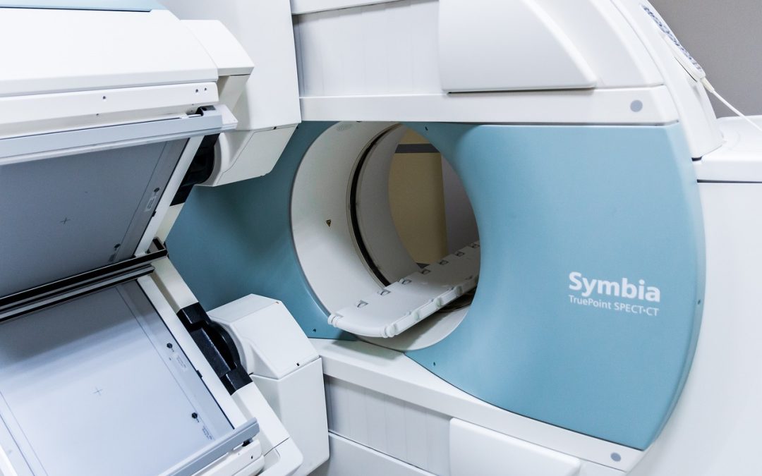
CT (computed tomography) scans are widely utilized in emergency situations where more detailed imaging is necessary. CT scans use a combination of X-rays and computer processing to create cross-sectional images of the body. They provide higher resolution images compared to traditional X-rays and are particularly effective in evaluating complex fractures, internal bleeding, or the presence of tumors. CT scans do involve higher radiation exposure than standard X-rays, but the benefits they offer in certain emergency situations often outweigh the associated risks.
Summary
In summary, X-rays play a critical role in emergency situations for various reasons. They assist in assessing fractures, identifying dislocations, detecting foreign objects, and evaluating joint or bone infections. X-rays provide quick and accurate diagnoses, guide treatment decisions, and minimize unnecessary procedures. The X-ray procedure during an emergency visit involves preparations, positioning, radiation protection, and image capture. While there are risks associated with X-rays, safety protocols, such as the use of lead aprons and limiting unnecessary exposure, help minimize these risks. Common concerns about X-rays include their effectiveness compared to other imaging techniques, costs, and minimizing radiation exposure. Determining the need for X-rays involves a clinical examination, healthcare provider’s assessment, and assessment of signs and symptoms. X-rays are useful for specific emergency conditions such as head injuries, chest and lung injuries, spinal cord injuries, and abdominal trauma. Alternatives to X-rays, such as ultrasound imaging, MRI scans, and CT scans, are available in certain cases. Overall, X-rays are commonly used in emergency situations, providing quick and accurate diagnoses, and safety protocols ensure the risks associated with X-rays are minimized.